THE SCIENCE
Signals within plasma cell-free DNA.
Cells shed fragments of DNA into the bloodstream. Their sequence, their physical characteristics and their epigenetic marks carry information about the tissue they came from.
A breakthrough already transforming human cancer care.
In human oncology, liquid biopsies analyse cell-free DNA in blood for molecular patterns associated with cancer.
Canos brings that scientific approach into veterinary medicine, combining genomic, physical and epigenetic information from canine cell-free DNA.
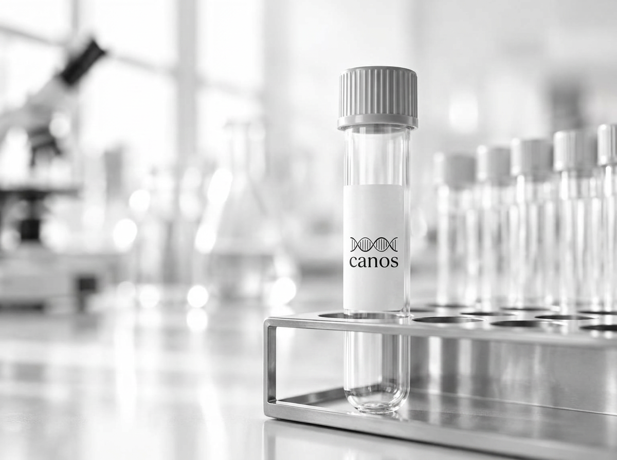
SPECIMEN · 5 ML · CELL-FREE DNA PRESERVATION TUBE
It starts with 5 mL of blood.
A routine blood draw collected by a veterinarian into a specialised preservation tube.
SCROLL
INSIDE THE SAMPLE
01 / 06
01 / 06
Separate
Plasma
The tube is spun. The plasma fraction is taken forward; the cellular fraction is not.
Cancer doesn’t leave one signal. It changes many.
Canos reads multiple properties of cell-free DNA together, including epigenetic patterns, fragment characteristics and genome-wide changes, to build a richer molecular picture.